
Figure 2. Proposed mechanism for TRα1PV/+ and mutant TRβPV mutant mice.
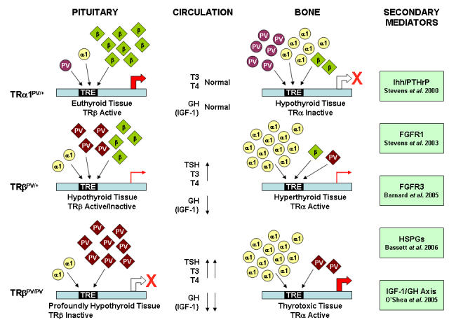
TRα1PV/+: In a TRβ predominant tissue, e.g. pituitary, the low levels of PV mutant receptor are unable to interfere with the actions of wild-type TRβ. In contrast, in a TRα1 predominant tissue, e.g. bone, the increased levels of dominant negative mutant receptor interfere with the actions of the wild-type receptors and impair the expression of T3-target genes. As a consequence, in TRα1PV/+ mutant mice, TRβ predominant tissues exhibit a euthyroid phenotype and TRα1 predominant tissues display a hypothyroid phenotype. TRβPV/+ and TRβPV/PV: In the TRβ predominant pituitary, there is a high level of TRβ expression relative to TRα1. Consequently, there are increasing levels of mutant and either low levels or no wild type TRβ in TRβPV/+ and TRβPV/PV tissues, respectively. In TRα1 predominant bone, the situation differs because levels of TRβ are low compared to TRα1. In both TRβPV/+ and TRβPV/PV mice, the low levels of mutant receptor are unable to interfere with the action of TRα1. Thus, in TRβPV mice, where the TRβ mutant acts as a rheostat and disrupts HPT axis regulation, TRβ predominant tissues display a hypothyroid phenotype with impaired expression of T3-target genes. TRα1 predominant tissues appear hyperthyroid in response to increased TRα1 activity that is stimulated by thyrotoxic circulating hormone levels resulting from impaired HPT axis regulation. The phenotype is less severe in TRβPV/+ heterozygous animals because peripheral hormone concentrations are less markedly elevated. A range of secondary mediators are included that have recently been shown to be targets of T3 action [Barnard et al., 2005; Bassett et al., 2006; O'Shea P et al., 2005; Stevens et al., 2003; Stevens et al., 2000].