
Fig. 7.
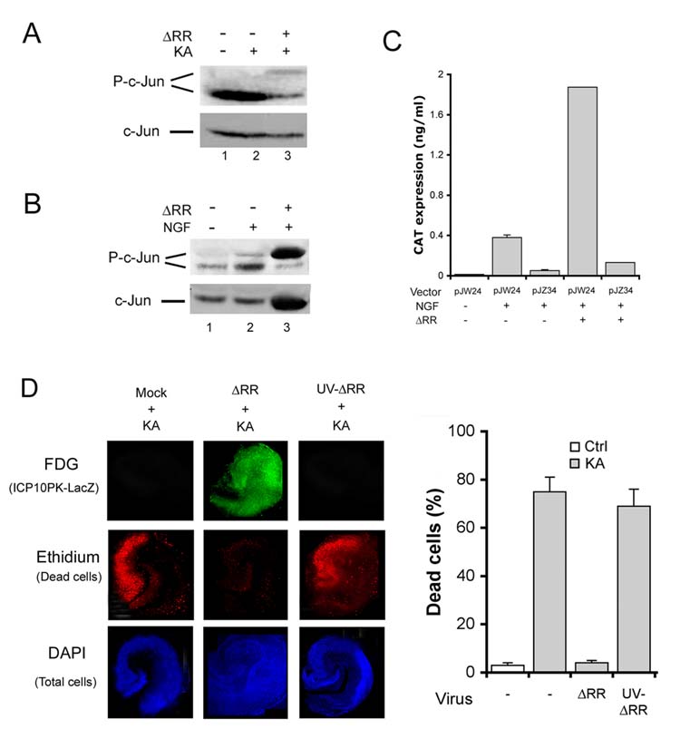
ICP10PK expression is regulated by an AP-1 feedback amplification loop. (A) Sprague Dawley rats were treated with ΔRR or PBS and given KA (15mg/kg; i.p.) at 24 hrs after the final treatment (day 0). Extracts of olfactory bulbs (day 8) were immunoblotted with P-c-Jun antibody. Blots were stripped and re-probed with antibody to total c-Jun. (B) Neuronally differentiated PC12 cells were infected with ΔRR (MOI=2) and extracts obtained at 8 hrs p.i. were immunoblotted with P-c-Jun followed by c-Jun antibodies. (C) Neuronally differentiated PC12 cells were transfected with pJW24 or pJZ34 (2 μg) using Fugene 6 transfection reagent and infected (or not) with ΔRR (MOI=2) at 24 hrs post transfection. CAT expression was measured by ELISA 24 hrs later and results are expressed as ng/ml (D). OHC treated (2hrs, 37°C) with ΔRR, UV-inactivated ΔRR (105 pfu), or mock treated with PBS and exposed to KA (5μM, 24 hrs) on day 2 were stained C12FDG (ICP10PK-LacZ expression) and ethidium homodimer (dead cells) and the number of ethidium homodimer staining cells counted as described in Materials and Methods. The % positive cells ± SEM was calculated relative to DAPI staining.