
Fig. 2.
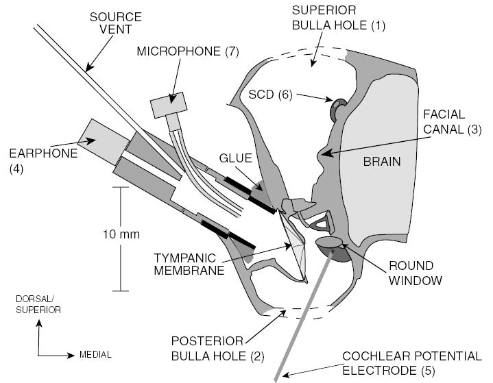
A schematic of the chinchilla middle-ear cavity (coronal view) illustrating the experimental setup. The superior hole (1) is opened to sever the facial nerve in its canal (3) and introduce the SCD (6). The posterior bulla hole (2) is opened to place the CP electrode (5) on the round window.