
Figure 4.
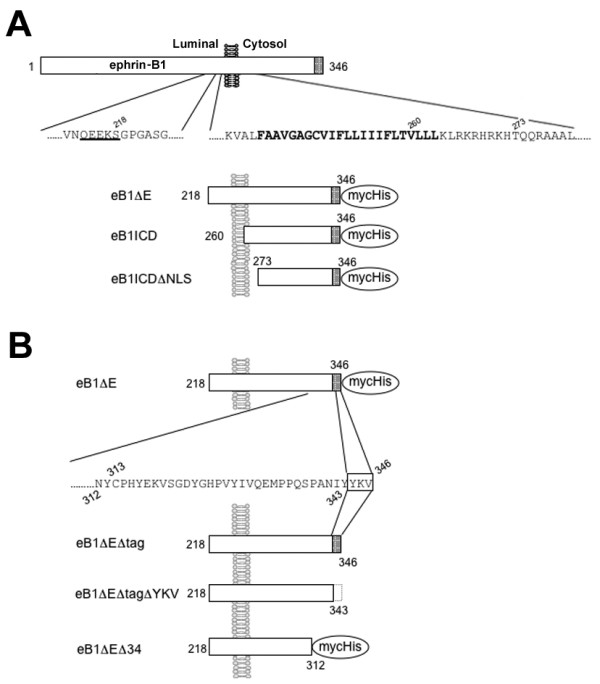
Schematic depiction of ephrin-B1 derivatives used in this study. A) N-terminally truncated ephrin-B1 derivatives. B) C-terminally truncated eB1ΔE derivatives. Putative amino acid sequence recognized by sheddase is underlined. Transmembrane domain is shown in bold. PDZ domain binding region (YKV) and Myc/His tag are indicated by shaded box and oval, respectively. Numbers indicate the residues in ephrin-B1 FL protein.