
Figure 1. Short-term evaluation of TEBV in canine model.
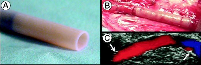
(a) Age- and risk-matched TEBV before implantation (4.2 mm ID) being removed from its temporary tubular support. (b) TEBV, anastomosed as end-to-end interpositional femoral graft, immediately after removal of the cross-clamps. (c) Doppler-ultrasound imaging at 2 weeks shows a patent vessel with uniform lumen and normal flow (arrows indicate anastomoses). TEBVs were endothelialized with autologous canine endothelial cells.