
Figure 2. Early remodeling of age- and risk-matched human TEBV after implantation in athymic rats.
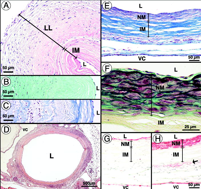
(a–c) Pre-implantation histology of TEBV (1.5 mm ID). (a) H&E staining reveals a decellularized internal membrane (IM), the living layer (LL) and the lumen (L) of TEBV. Lumens of vessel were seeded with syngeneic rat endothelial cells. Note that the wall is scalloped because this vessel was an unused surgical sample that contracted prior to fixation. (b) Movat staining reveals the large proteoglycan (aqua green) content of the TEBV at the time of implantation. (c) Verhoff-Masson staining indicates the high collagen (blue) content of the TEBV. (d) 90 days after implantation, H&E staining of the perfusion-fixed graft shows complete tissue integration with minimal inflammatory/immune response and a modest neomedia formation. VC: vena cava. (e) Verhoff-Masson staining clearly indicates that the IM was largely 16 intact and still acellular. The neomedia was rich in collagen and cells and appeared to have elastic fibers (black). (f) At higher magnification, a Movat staining reveals the forming elastic fibers and a developing internal elastic lamella-like structure forming under a confluent endothelium. Note that proteoglycans were present in the neomedia and subendothelial space but not no longer in the IM (yellowish staining indicates collagen). (g–h) Immunohistochemical staining for von Willebrand factor and smooth muscle α-actin reveals the confluent endothelium and the SMC layers (respectively) of the TEBV and adjacent vena cava. Note the presence of SMC-specific α-actin positive cells at the IM/adventitia interface.