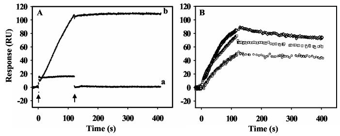
Figure 1.
SPR sensorgrams showing proline and ligand induced PutA-lipid binding. Panel A: Sensorgrams of PutA (20 nM) in the oxidized state (a) and in the presence of 5 mM proline (b) binding to a lipid bilayer of E. coli lipid polar extracts. The arrows indicate the beginning and end of the protein sample injection. In sensorgram a, the rapid change in response units (RU) at the injection start point is due to a refractive index change of the sample, not by protein binding to the lipid bilayer. The RU returned to the initial value after the injection of the sample was complete. Panel B: Oxidized PutA (80 nM) in the presence of 5 mM L-THFA ( ○ ), 5 mM L-lactate ( □ ) and 5 mM DL-P5C ( ◇ ) was injected at 60 μl/min for 120 s onto a L1 chip coated with E. coli polar lipid vesicles. The dissociation phase was observed by the flow of HEPES-N buffer at 60 μl/min for 300 s.

