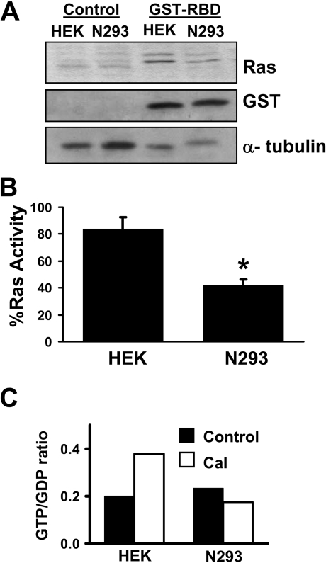
Figure 3. NO• inhibits H-Ras activity.
(A) HEK-293 (HEK) or N293 cells were treated with 10 μM A23187 for 2 h. Cell lysates were subjected to the H-Ras pull-down assay using GST–RBD. GST–RBD protein complexes were separated by SDS/PAGE (15% gels) and immunoblotted for H-Ras or GST. An anti-α-tubulin immunoblot from the initial lysates was included as a protein loading control. (B) The relative amount of active H-Ras co-interacting with GST–RBD was quantified by densitometry. Results are means±S.D. for three independent experiments. * P<0.05. (C) HEK-293 (HEK) and N293 cells were incubated in DMEM medium containing 0.5 mCi of carrier-free [32P]Pi for 4 h. The cells were then stimulated with 10 μM A23187 for 1 h, lysed and then immunoprecipitated for 2 h at 4 °C with anti-H-Ras antibody. H-Ras-bound nucleotides (GTP and GDP) were eluted and separated by TLC. GTP/GDP ratios were quantified by PhosphorImaging. The data represent the means for two independent experiments. The GTP/GDP ratios for the two experiments were 0.13 and 0.27 for HEK-293 control cells; 0.45 and 0.31 for HEK-293 cells plus A23187 (labelled Cal); 0.21 and 0.26 for N293 cells; and, 0.17 and 0.18 for N293 cells plus A23187 (labelled Cal).

