Fig. 6.

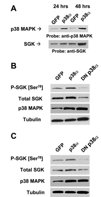
Correlation between p38α expression and SGK. A: KMCH cells were infected with 5 × 10⁶ PFU/ml adenoviral (Adv)-p38α or Adv-green fluorescent protein (GFP) (controls). Nuclear extracts were prepared after 24 or 48 h, and immunoblot analysis was performed using specific antibodies to p38 MAPK or SGK. KMCH cells (B) or Mz-ChA-1 cells (C) were infected with Adv constructs encoding GFP, p38α, or DNp38α. Whole cell lysates were prepared 48 h after infection and assessed by performing immunoblot analysis using a Ser78 phospho-specific SGK antibody. The blots were stripped and reprobed with phosphorylation state-independent antibodies to SGK (total SGK), p38 MAPK, and α-tubulin (loading control).
