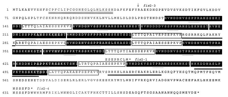
Figure 2.
Deduced protein sequence of FIS2. The amino acid sequence corresponding to the C2H2 zinc finger motif is double underlined. The three putative nuclear localization signals are underlined with thick lines. The 12 A repeats are shown in filled boxes and the 7 B repeats are shown in open boxes. The [T/S]PXX motifs within the A repeats are underlined with thin lines. The position of the fis2–1 (deletion of T) and the fis2–4 (G → A) mutations are shown with the resulting modifications of the encoded peptide. The location of the fis2–3 mutation (G → A) at the junction of intron 5 and exon 6 is indicated by an arrow (↓). Stop codons are indicated with asterisks (∗).

