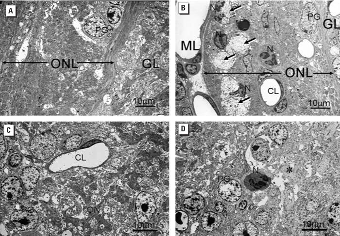
Figure 9.
Ultrastructural alterations in ONL and GL of OB 7 days PI with saline vehicle alone (A, C) or SG (500 μg/kg bw) (B, D). Abbreviations: CL, capillary lumen; GL, glomerular layer; ML, meningeal layer; N, neutrophil; ON, olfactory nerves; ONL, olfactory nerve layer; PG, periglomerular cell. (A, B) Focal areas of axonal degeneration (long arrows) present in the markedly atrophic ONL in the SG-treated mouse (B) but not the vehicle-treated control mouse (A). In (B), infiltrating Ns with segmented nuclei are closely associated with areas of degeneration (arrows). (C, D) Dense staining axons of ON in the GL of the vehicle control mouse (C) are absent in the GL of the SG-exposed mouse (asterisk in D). An N is also present in the SG-exposed mouse but not in the control mouse.

