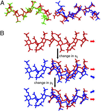
Figure 2.
Kink in FGF-bound oligosaccharides. The direction of the chains shown in A and B is from nonreducing end to reducing end (left to right). (A) Superimposition of FGF-1-bound (green) and modeled unbound oligosaccharide (on the left) and FGF-2-bound (blue) and modeled unbound oligosaccharide (on the right). The ring atoms and the glycosidic oxygens (O1 and O4) of H3 (Fig. 1A) in bound and unbound oligosaccharide structures were superimposed. H3-I4-H5 (Fig. 1A) of FGF-1-bound oligosaccharide and H1-I2-H3 (Fig. 1A) of FGF-2-bound oligosaccharide constitute the trisaccharide-spanning kink. (B) Stepwise formation (top to bottom) of the kink and continuation of helical structure caused by changes in p1j and s1j (according to the boldface values in Table 1) of the oligosaccharide (blue) relative to the unbound oligosaccharide (red).

