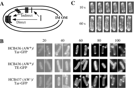
Fig. 1. Polar localization of Tar–GFP.
A. Direct and indirect membrane insertion models for polar localization of the chemoreceptors. To target to a cell pole, a nascent chemoreceptor protein might be inserted into the cytoplasmic membrane (i) at or near a cell pole (a direct model), or (ii) at random positions thereafter migrating or diffusing away from the insertion point (an indirect model). IM, the cytoplasmic (inner) membrane; OM, the outer membrane.
B. Time-course of polar localization of Tar–GFP. HCB436 (CheAW+) or HCB437 (CheAW−) cells carrying a plasmid encoding Tar–GFP or Taz1–GFP were observed at indicated time points after the addition of 1 mM arabinose. Numbers above pictures represent harvested time points (min).
C. Time-lapse observation of polar localization of Tar–GFP. HCB436 cells carrying plasmid encoding Tar–GFP were spotted onto a glass slide covered with 0.5% agarose. Fluorescence was observed with 10 or 60 s intervals.

