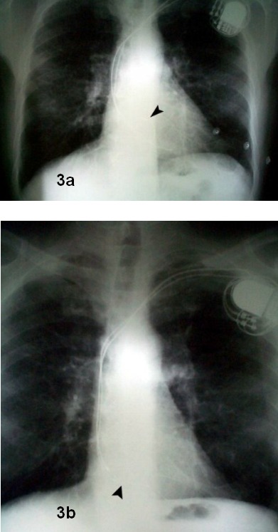
Figure 3.

Posteroanterior chest x-ray film. Top (A): radiograph obtained 24 hours after pacemaker implantation. Arrowhead shows atrial lead tip inside the right atrial appendage. Bottom (B): radiograph obtained three months later, showing displacement of atrial lead towards tricuspid annulus. However, diagnosis is not evident from the posteroanterior view alone, due to superimposition of structures and radiograph densities.
