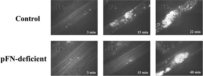
Figure 2.
Thrombus growth in the presence or absence of pFN. Times after FeCl3-induced injury are indicated in bold. In the control mouse (Upper) and the pFN-deficient mouse (Lower), single fluorescent platelets are seen to adhere in the arterioles at 3 min after injury. Whereas in the control vessel large stable thrombi grew (15 min), leading to occlusion at 22 min, only very small thrombi were apparent at 15 min in the pFN-deficient animal, and at termination of the experiment (40 min) the vessel was still patent. The lack of stability of the thrombus formed in the pFN-deficient mouse is better observed in Movie 1, where the two arterial segments are shown in real time.

