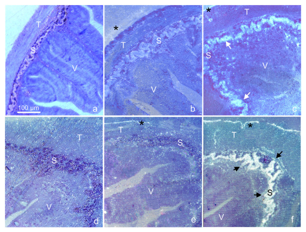
Figure 2.

PrPSc accumulation in the pyloric caeca (a, b, c) and non-everted intestine (d, e, f) of trout intestine statically perfused. Immunohistochemistry on trout sections of pyloric caeca (a, b, c) and non-everted intestines (d, e, f). The static perfusion was performed for 1 hour at 15°C, in a PBS solution containing 50 μl/ml of either 10% uninfected mice brain homogenate (a, d) or 10% mice scrapie brain homogenate (139A) (b, c, e, f). Immunolabelling was performed with the monoclonal antibody SAF83 (a, c, d, f), or, as control, the monoclonal anti-HA against influenza virus (anti-HA, clone 12CA5, subtype IgG2b, k), (b, e). All the immunogold-labelled sections were silver enhanced and counterstained with 0.1% toluidine blue (see materials and methods for details). PrPSc localisation (arrows) occurred in the stratum compactum (S) of distal intestine and pyloric caecum incubated with SAF83 (c, f). No immunolabelling was present in control tissues incubated with the same antibody (a, d), except for a low unspecific background. The background was slightly higher, though unspecific, in anti-HA labelled sections (b, e) due to the high reactivity of this antibody. All micrographs were taken at the same magnifications; V = villo, T = tunica muscolaris; asterisks point to unspecific labelling present in all samples and due to micro-fractures between the outer specimen surface and the resin.
