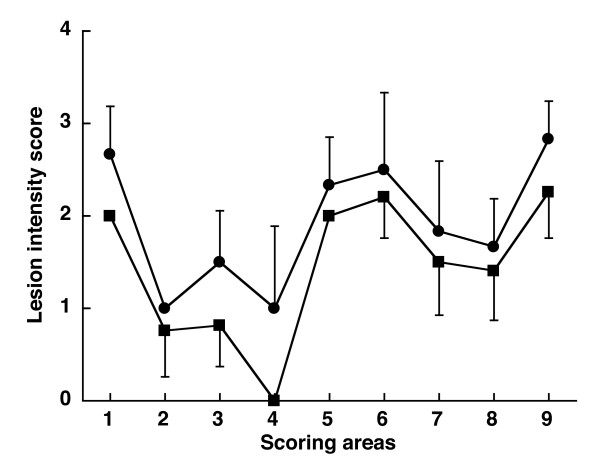
Figure 3.

Lesion profiles for mice following inoculation with 139A before (circle) and after (square) passage in fish. The lesion profiles for the fish-passaged 139A (square) was a mean of 4 mice: 2 mice inoculated with turbot spleen at 15 days, 1 mouse inoculated with trout spleen at 15 days, and 1 turbot brain taken 90 days after parenteral inoculation. The reference curve (circle) was a mean of 6 mice inoculated with the 139A non passaged in fish. Vacuolation was evaluated in nine standard areas: 1, dorsal medulla; 2, cerebellar cortex; 3, superior colliculus; 4, hypothalamus; 5, thalamus; 6, hippocampus; 7, septum; 8, retrosplenial and adjacent motor cortex; 9, cingulate and adjacent motor cortex. Data are mean ± SE.
