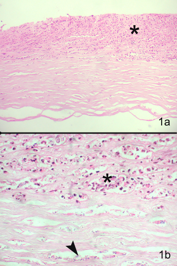
Figure 1.

Section of the corneal tissue shows epithelial ulceration, with inflammatory infiltrates in the anterior two-thirds of stroma (hematoxylin & eosin, × 1100) (b) Higher magnification shows polymorphonuclear cells (asterix) and faintly stained, ill-defined oval dot like structures (arrow) between the corneal lamellae (Hematoxylin and Eosin stain, × 400)
