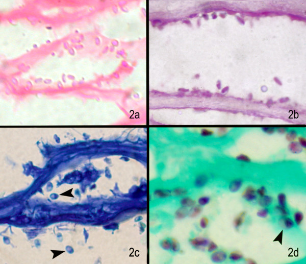
Figure 2.

(a) Under higher magnification, the microsporidal spores are seen as pink oval structures (H & E stain, × 1000); (b) magenta pink oval structures in PAS stain (× 500), (c) deep blue oval structures with dark tip (arrow) in some spores Giemsa stain (× 500), (d) well defined brown oval spores with dark tip or band in Gomoris methenamine silver stain (× 500)
