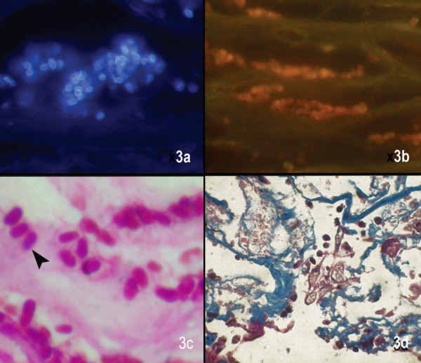
Figure 3.

Microsporidal spores are seen as white fluorescent ring like structures in Calcofluor white stain (× 500), (b) dull oval orangish structures in Acridine orange stain (× 500), (c) oval well defined spores with a faint hollow around the spore in Gram stain (× 1000), (d) dark blue uniformely stained bodies in Masson's trichrome stain (× 500)
