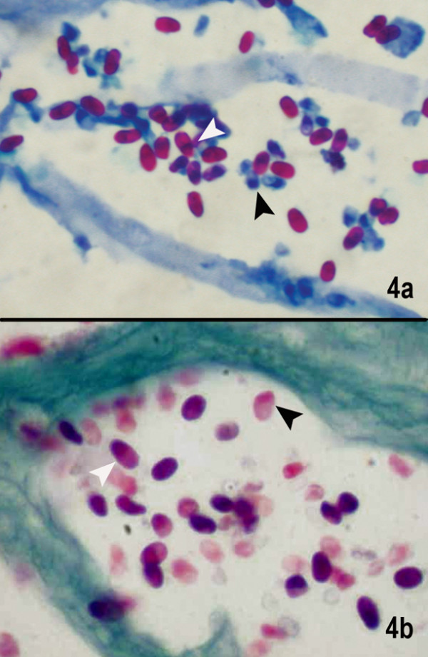
Figure 4.

(a) Microsporidal spores are seen as well defined oval reddish bodies with a dark staining of the narrow end of the spore (black) or a waistband (white) closer to the tip of narrow end. Also seen are the unstained blue spores which possibly are immature or degenerating spores (1% acid fast stain, × 1000).(b) The spores are well delineated as purplish pink egg-shaped spores with a darker staining of the tip (white arrow). Even the degenerating spores show the darkly staining tip (black arrow) Gram's chromotrope stain (× 1000)
