Figure 5.

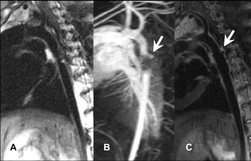
Early detection of aortic perforation. A, DIR-FSE image of coarctation at before stenting. B, Contrast exit at superior edge of stent (arrow). C, DIR-FSE shows apposed oversized stent with blood accumulation along aortic wall (arrow).

Early detection of aortic perforation. A, DIR-FSE image of coarctation at before stenting. B, Contrast exit at superior edge of stent (arrow). C, DIR-FSE shows apposed oversized stent with blood accumulation along aortic wall (arrow).