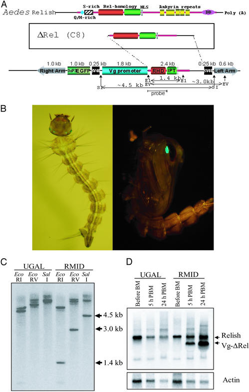
Figure 1.
Structure of the pBac[3xP3-EGFP, afm, Vg-ΔRel] transformation vector and its expression in transgenic mosquitoes. (A) Schematic diagram of the pBac[3xP3-EGFP, afm, Vg-ΔRel] transformation vector that was transformed into the A. aegypti germ line. (B) Detection of the transgenic Vg-ΔRel mosquitoes by transformation marker-mediated fluorescence. (C) Southern blot analysis of genomic DNA extracted from the Vg-ΔRel transgenic mosquitoes and the parental UGAL strain, digested with EcoRI, EcoRV, and SalI. The probe region and the restriction sites of the transformation vector are indicated in A. (D) The expression profile of Relish and transgenic Vg-ΔRel transcripts after blood feeding. Endogenous ≈3.9-kb Relish transcript was detected in both transgenic and parental mosquitoes at any stage. An additional band of ≈3.0 kb was detected in the transgenic mosquitoes; it appeared after bloodfeeding and reached its peak at 24 h PBM.

