
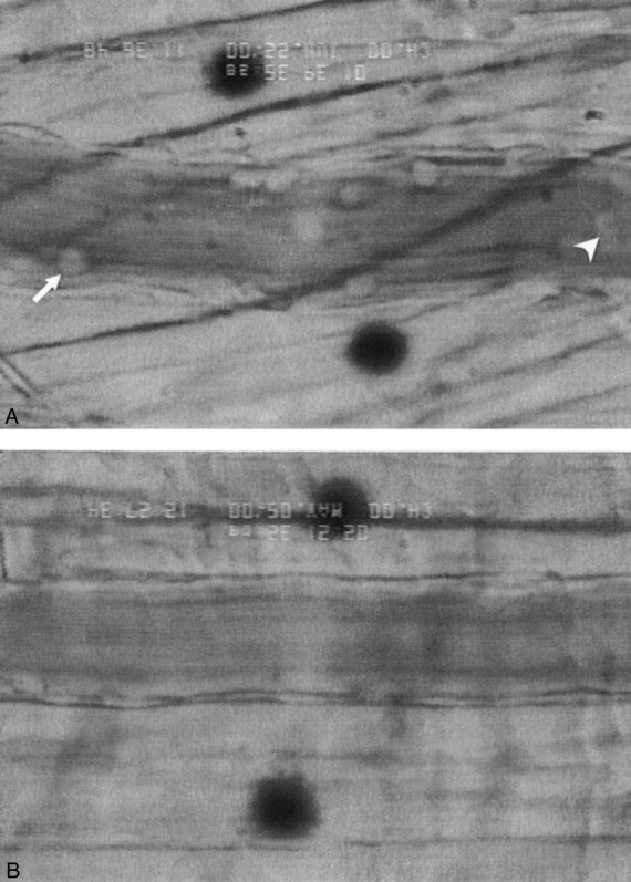
Figure 6. Representative examples of intravital microscopic images containging the lack of PMN rolling (arrowhead) and PMN adherence (arrow) in HTS (A) as compared to RL (B) vessels 90 minutes after resuscitation.
Official websites use .gov
A
.gov website belongs to an official
government organization in the United States.
Secure .gov websites use HTTPS
A lock (
) or https:// means you've safely
connected to the .gov website. Share sensitive
information only on official, secure websites.
Figure 6. Representative examples of intravital microscopic images containging the lack of PMN rolling (arrowhead) and PMN adherence (arrow) in HTS (A) as compared to RL (B) vessels 90 minutes after resuscitation.