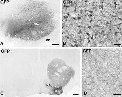
Figure 1.
Photomicrographs showing the expression of the GFP transgene in monkeys injected with the control vector. (A and B) GFP was expressed in the vast majority of the neurons in SN pars compacta, and some cells dorsally in the mesephalic tegmentum and ventrally in the SN pars reticulata (A). The transgenic GFP protein was transported along the axons of the nigrostriatal projection system to the caudate nucleus, putamen, and lateral olfactory tubercle, whereas the projections to the medial olfactory tubercle and the nc. accumbens were not labeled to the same extent (C; the noninjected control side is to the left). The density of GFP-expressing fibers in the striatum is shown in D. Scale bar in A = 0.5 mm; B and D = 50; C = 1 mm.

