Figure 3.
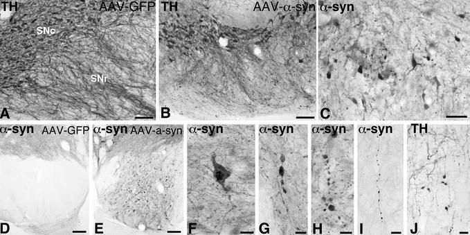
Nigral degeneration in the rAAV-α-syn-injected animals. Pathological profiles in the SN were seen both with TH (A, B, and J) and α-syn immunoreactivity (C–I). Extensive cellular degeneration in the SN pars compacta, as visualized by TH immunoreactivity, also led to prominent dendritic abnormalities and loss of dendrites in the SN pars reticulata (B and J). Affected but surviving cells in SN pars compacta were shrunken (C) and contained numerous inclusions within the cytoplasm and proximal neurites (F), as well as in axons within pars compacta (G and H) and along the nigrostriatal bundle (I). Numerous pathological fibers with prominent inclusions were observed in the cerebral puduncle (cf. D and E). Scale bars in A and B = 0.1 mm; C, I, and J = 50 μm; D and E = 0.2 mm; F, G, and H = 25 μm.

