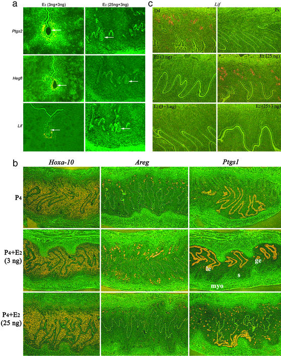
Figure 2.
Uterine gene expression in receptive and refractory uteri. (a) Lif, Ptgs2, and Hegfl expression is aberrant at the site of a blastocyst in a refractory uterus induced by 25 ng of E2. Recipient mice were ovariectomized on day 4 of pseudopregnancy and injected daily with 2 mg of P4 to induce the condition of delayed implantation. On day 7, the recipients received the first injection of E2 at 3 or 25 ng. On day 8, blastocysts were transferred into these recipients immediately followed by a second injection of 3 ng of E2. Uterine sections containing blastocysts were processed for in situ hybridization 24 h later. (Magnification, ×100.) Arrows indicate the location of blastocysts. (b) Uterine expression of Areg and Ptgs1 becomes aberrant at a higher E2 level. Pseudopregnant mice ovariectomized on day 4 were injected daily with P4 to induce the condition of delayed implantation. On day 7, they received an injection of 3 or 25 ng of E2. Uteri were processed for in situ hybridization 24 h later. (Magnification, ×40.) Note that although Hoxa-10 expression is similar at 3 or 25 ng of E2, the expression of amphiregulin and COX-1 is aberrant at 25 ng of E2. le, luminal epithelium; ge, glandular epithelium; s, stroma; myo, myometrium. (c) Uterine Lif expression is different at higher and lower doses of E2. As stated above, pseudopregnant mice ovariectomized on day 4 were treated with P4 daily to induce the condition of delayed implantation. On day 7, they received 3 or 25 ng of E2 or received the first injection of E2 at 3 or 25 ng followed by a second injection of 3 ng of E2 24 h later. Uteri were processed for in situ hybridization at different times. Results of Lif expression at 24 h are shown. (Magnification, ×100.) E2 at 3 ng as one or two injections failed to detect glandular Lif expression, whereas 25 ng of E2 as a single injection induced this expression. The expression was undetectable when the first E2 injection was at 25 ng followed by a second injection at 3 ng. Representative sections of day 4 (D4) pregnant uterus showing glandular Lif expression and of P4-treated uteri showing the absence of Lif expression were used as positive and negative controls, respectively.

