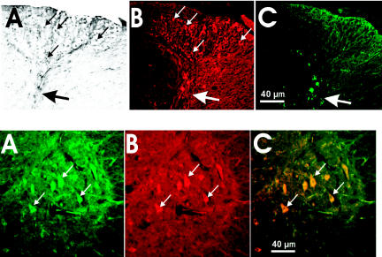
Figure 10.
Survival of ES cells ipsilateral to the lesion at 60 days. Upper panel: Immunodetection of GFP (A, DAB staining), NeuN (B, immunofluorescence), and GFAP (C, immunofluorescence) in the ipsilateral dorsal horn. Small white or black arrows indicate neurons. Big white or black arrow points at collapsed dorsal horn. Lower panel: Double immunofluorescence for GFP and MAP-2 (a neuronal marker). (A) GFP fluorescent cells. (B) MAP-2–positive cells. (C) Merge of 2 images. Small white arrows indicate neurons.

