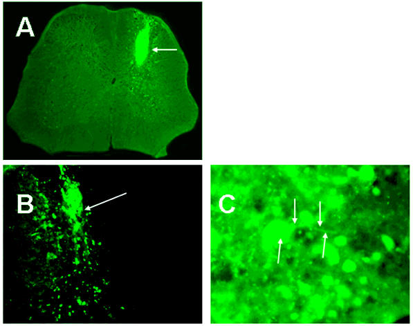
Figure 7.
Fluorescence microscopy of GFP-expressing cells 14 days after ES cell transplantation (A–C). (A) GFP cells fill epicenter of injury. Arrow indicates transplanted cells. (B) GFP cells migrating into surrounding tissue. Arrow indicates origin of migrating GFP-positive cells. (C) GFP cell with processes. Arrows indicate cell body and processes.

