Figure 6.
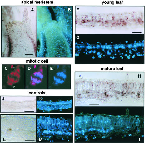
Immunolocalization of GRIMP.
N. benthamiana tissue sections were analyzed using anti-GRIMP IgG ([A] to [I]) or preimmune IgG ([J] to [M]) followed by anti-rabbit secondary antibody and peroxidase staining ([A], [B], and [F] to [M]) or Texas red–conjugated secondary antibody ([C] to [E]). All sections also were stained with DAPI. Bars = 50 μm.
(A) and (B) Meristem showing GRIMP (A) and DAPI (B) staining.
(C) to (E) Confocal image of a mitotic cell treated with anti-GRIMP IgG and a Texas red–labeled secondary antibody (C), DAPI staining (E), and the merged image (D). Open arrowheads show GRIMP staining associated with the spindle poles.
(F) and (G) Young leaf showing GRIMP (F) and DAPI (G) staining.
(H) and (I) Mature leaf showing GRIMP (H) and DAPI (I) staining.
(J) and (K) Young leaf treated with preimmune IgG showing no peroxidase staining (J) but stained with DAPI (K).
(L) and (M) Mature leaf treated with preimmune IgG showing no peroxidase staining (L) but stained with DAPI (M).

