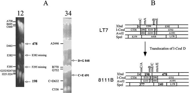
FIG. 3.
Genomic translocation exemplified by strain 8111B. (A) PFGE gels of LT7 and 8111B. Lanes: 1, LT7 DNA cleaved by XbaI, with the fragments and their sizes indicated on the left of the PFGE gel; 2, 8111B DNA cleaved by XbaI, with deviations of the cleavage pattern from that of LT7 indicated on the right of the PFGE gel; 3, LT7 DNA cleaved by I-CeuI, with the fragments and their sizes indicated on the left of the PFGE gel; and 4, 8111B DNA cleaved by I-CeuI, with deviations of the cleavage pattern from that of LT7 indicated on the right of the PFGE gel. (B) Local comparison of LT7 and 8111B showing the translocation of I-CeuI D, which resulted in three hybrid rrn operons, disappearance of two XbaI fragments (F and E) and two SpeI fragments (K and A [not shown on the PFGE picture]), and appearance of two new fragments each from XbaI (198 and 478 kb) and SpeI (377 and 240 kb) digestions.

