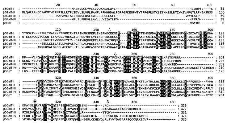
Figure 2.
clustalw alignment of mouse β3GalT-I, -II, -III, and -IV and mouse β3GnT proteins. Conserved residues are shaded. The white arrows mark the positions of the cysteine residues conserved among β3GalT proteins. The black arrow shows the position of the cysteines conserved in the five proteins.

