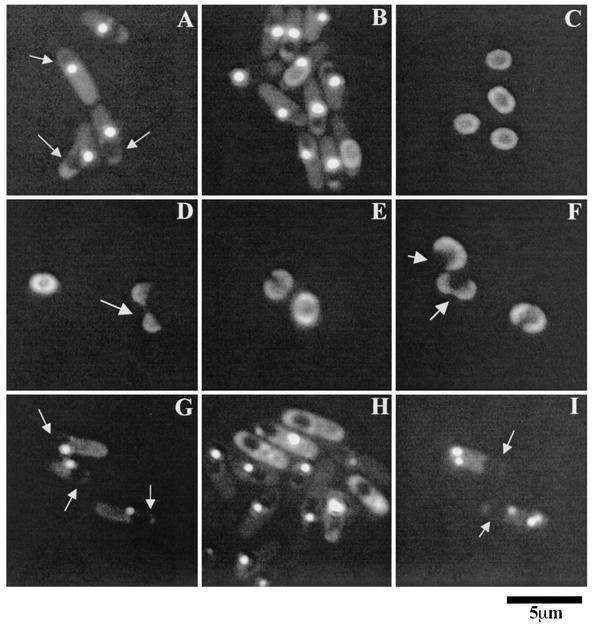
FIG. 6.
Localization of YwdL-GFP in sporulating cells and dormant spores. (A to C) Strain KB48 (ywdL-gfp), was sporulated by resuspension, and samples were viewed with a fluorescence microscope at 3 h (A), 7 h (B), and 24 h (C) after the onset of sporulation. (D to F) Dormant spores of strain KB48 were germinated in nutrients at 37°C for 1 h, and samples were examined. Arrows indicate germinated spores whose coats have split open. (G to I) Strains KB62 (ΔcotE ywdL-gfp) (G and H) and CVO1736 (ΔspoIVA ywdL-gfp) (I) were sporulated by resuspension, and samples were viewed at 3 h (G and I) and 7 h (H) after the onset of sporulation. Strain KB59 released dormant spores with no visible YwdL-GFP on their periphery, while strain CVO1736 did not release any dormant spores. The arrows pointing to the dark areas enclosed in the sporulating cell in panels A, G, and I indicate the developing forespore.

