Figure 1.
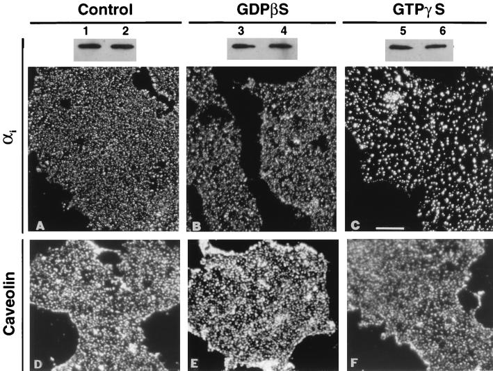
GTPγS alters the immunofluorescence pattern for αi but does not release the protein from the plasma membrane. MA104 cells were grown on coverslips and then sonicated to isolate fragments of adherent plasma membrane with their inner surface exposed. Membranes were processed for immunofluorescence (A–F), or duplicate samples were solubilized in SDS/PAGE sample buffer and processed for Western immunoblotting (lanes 1–6). Membranes, viewed en face in A–F, were either processed immediately (control, A and D, lanes 1 and 2) or incubated at 37°C for 30 min in the presence of 10 μM guanosine 5′-(2-O-thio)diphosphate (GDPβS; B and E, lanes 3 and 4) or GTPγS (C and F, lanes 5 and 6) before processing. Affinity-purified B087 antibodies (specific for αi) were used at a concentration of 10 μg/ml for immunofluorescence (A–C) and 50 ng/ml for immunoblotting (lanes 1–6). Antibodies against caveolin were diluted to 1 μg/ml (D–F). Texas Red conjugated to goat anti-rabbit IgG (20 μg/ml) was used as the secondary antibody in A–F. (Bar = 4 μm.)

