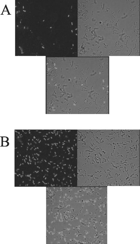
FIG. 2.
Fluorescence microscopy of Sytox Green-stained cells with and without induction of Fst. Cells were cultured in N2GT medium for 1 h and then induced with either cAD1 (100 ng/ml) or an equal volume of dimethyl sulfoxide for another hour. Cells were harvested, washed, and stained with Sytox Green as described in Materials and Methods. For fluorescence microscopy in A and B, wet mounts were prepared and examined under oil with a 100× objective. For each panel, three views are shown: fluorescence alone (upper left), transmitted light alone (upper right), and an overlay of both (bottom). A, uninduced; B, induced.

