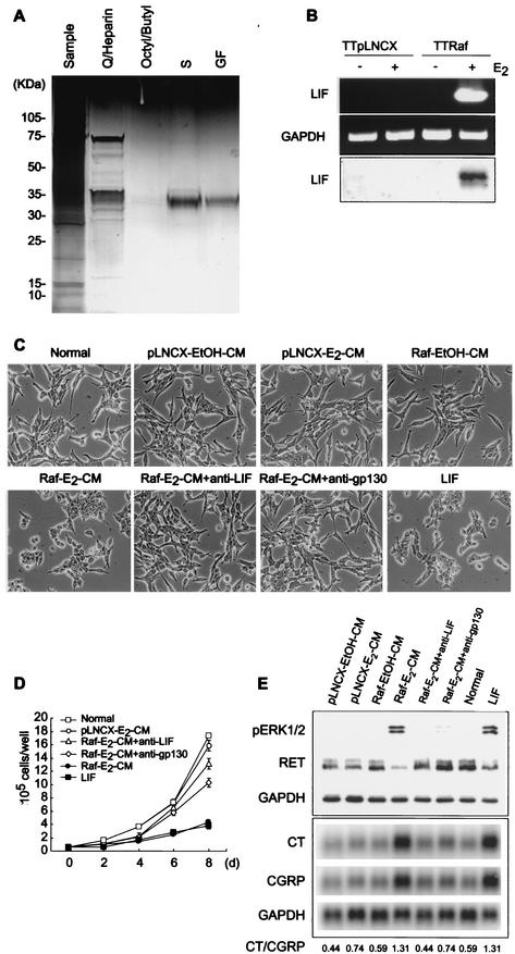
FIG.2.
LIF is the autocrine growth inhibitor induced by Raf. (A) Conditioned medium was concentrated by ultrafiltration, dialyzed (sample), and subjected to a series of protein purification columns, consisting of the anion exchanger unoQ6 coupled with heparin-Sepharose (Q/Heparin), octyl-Sepharose coupled with butyl-Sepharose (Octyl/Butyl; flow through), the cation exchanger unoS1 (S), and Superdex G200 gel filtration (GF). Active fractions from each column were resolved by SDS-PAGE and visualized by silver staining. (B) Expression of LIF upon Raf activation was investigated by RT-PCR of total RNA from TTpLNCX or TTRaf cells treated with ethanol or estradiol (top and middle panels) and by Western blot detection of LIF in cell culture media (bottom panel). (C to E) TT cells were treated with the control conditioned media, Raf-E2-CM, LIF-neutralized Raf-E2-CM (Raf-E2-CM+anti-LIF), and LIF. Cells were also pretreated with anti-gp130 blocking antibody prior to Raf-E2-CM treatment (Raf-E2-CM+anti-gp130). Cells were then observed for changes in morphology (C), growth (D), and expression of phosphorylated ERK1/2, RET (Western hybridization), or calcitonin genes (Northern hybridization) after 2 days (E). Data (mean ± standard error) are from a representative experiment performed in triplicate. P value is <0.001 for Raf-E2-CM+anti-LIF and Raf-E2-CM+anti-gp130 compared to Raf-E2-CM on cell growth curve (one-way analysis of variance). Experiments were repeated at least three times with similar results.

