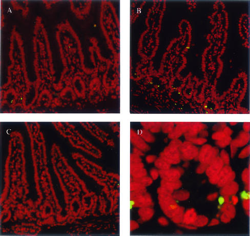
FIG. 5.
Increased number of apoptotic cells in crypts of small intestines from mdm2puro/Δ7-12 mice. TUNEL staining (green) was performed on 5-μm sections of the small intestine. Propidium iodide (red) was used to stain all nuclei. (A) Wild type. (B) mdm2puro/Δ7-12. (C) mdm2puro/Δ7-12 p53−/−. (D) Close-up of crypt from mdm2puro/Δ7-12 mouse small intestine.

