FIG. 2.
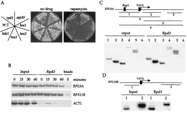
Rpd3 localizes at RP genes independently of Tor signaling or RP gene activators. (A) Isogenic wild-type (S288C), and hos1, hos2, hos3, hda1, rpd3, and sap30 mutant strains were grown on YEPD (no drug) or YEPD containing 100 ng of rapamycin ml−1. After 3 days of incubation at 30°C the plates were photographed. (B) Exponentially growing cultures of strain MCY47 were treated with 100 nM rapamycin for 0, 15, 30, or 60 min and cross-linked with formaldehyde. Chromatin was prepared and immunoprecipitated with antibodies specific for Rpd3. PCR was performed in the total chromatin (Input) or the immunoprecipitated DNA with specific primers for the RPL9A, RPS11B, and ACT1 gene promoters indicated on the right. Controls were performed with protein G beads. The results shown are representative of two independent experiments. (C and D) The Rpd3 immunoprecipitated DNA from rapamycin-untreated cells in the experiment shown in Fig. 2B was amplified with primers specific for RPL9A (panel C) or RPL18B (panel D). The PCR products encode the following sequences: RPL9A, −398 to −98 (1), 35 to 300 (2), 917 to 1154 (3), −398 to 300 (4), 35 to 1154 (5), −398 to 1154 (6) in RPL18B, −410 to −151 (1) and 1216 to 1500 (2). For a control, we performed PCR with the DNA obtained from the mock immunoprecipitation (with no antibody but beads from the same experiment) and the different sets of primers. The results obtained were very similar to those of the beads control presented in Fig. 2B.

