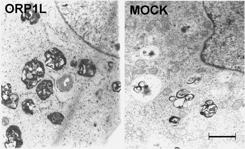
Figure 6.
Electron microscopy of endosome morphology in cells stably transfected with ORP1L cDNA (ORP1L) or the empty vector pcDNA3.1 (MOCK). The cells were fixed with glutaraldehyde, processed for electron microscopy using a standard procedure, and horizontal sections were viewed with an EX200 electron microscope (JOEL). The ORP1L-expressing cells display abundant multivesicular body/late endosome-like organelles that are abnormally full of electron dense internal membranes. Bar, 1 μm.

