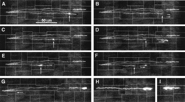
Figure 3.
(A) A chromosomal fragment (≈870 kbp) is trapped at 10 V/cm, its lower arm is successively dissected with an argon–ion laser beam (200 mW, 488 nm), and the excised DNA fragments migrate away from the field of view (B–D). Eventually, a point is reached where the molecule starts slipping off the U-shape (F–I).

