Figure 5.
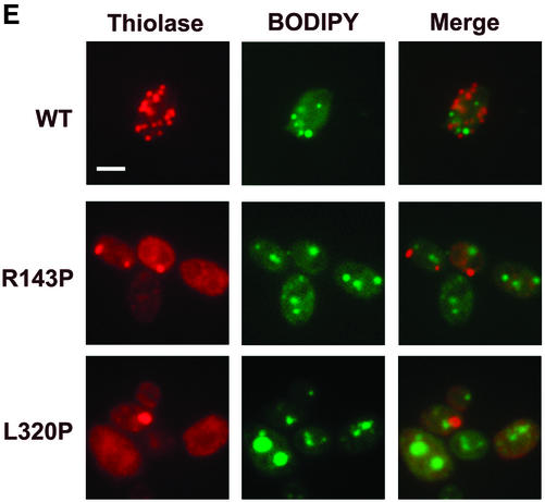
De novo peroxisome biogenesis occurs in cells grown in glucose-containing medium in regions associated with ER elements. (A) Immunofluorescence analysis demonstrating that the peroxisome-like structures found at the periphery of glucose-grown MycPex3p-R143p– or MycPex3p-L320P–expressing cells contain the matrix enzyme AOX but not the matrix enzyme thiolase, suggesting that these structures are nascent peroxisomes undergoing biogenesis. Bar, 3 μm. (B) Peripherally located peroxisomes contain different matrix proteins. Y. lipolytica cells were analyzed by immunofluorescence with antibodies to thiolase, the PTS1 SKL, AOX, and ICL. Bar, 3 μm. (C) Nascent, peripherally located peroxisomes are found in ER-rich regions of the cell and sometimes associated with the nuclear envelope. Cells were processed for immunofluorescence and doubly labeled with antibodies to thiolase and to the ER resident protein Kar2p. Bar, 3 μm. (D) Individual, peripherally located peroxisomes are often positioned close to nuclei. Cells were labeled with antibodies to thiolase and with the nuclear stain, DAPI. Bar, 3 μm. (E) Peroxisomes are randomly positioned with respect to lipid droplets in glucose-grown cells. Cells were labeled with antibodies to thiolase and with the neutral lipid stain BODIPY 493/503. Bar, 3 μm. (F) Peroxisome-like structures (P) are observed by electron microscopy closely associated with ER elements (E) in cells undergoing de novo peroxisome biogenesis. Cells were grown in glucose-containing YND medium at 16°C for 48 h.


