Figure 8.
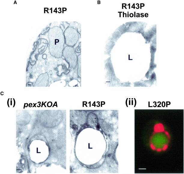
Microscopic analysis of peroxisomes in cells expressing MycPex3p-R143P and of structures surrounding lipid droplets. Cells were grown for 48 h in glucose-containing YND medium at 16°C, transferred to oleic acid-containing YNO medium, and incubated for a further 8 h at 16°C. (A) Electron micrograph of peroxisomes (P) in MycPex3p-R143P–expressing cells. (B) Electron micrograph of the structure surrounding a lipid droplet (L) in cells expressing MycPex3p-R143P. The structure shows immunogold labeling with antibody to thiolase. (C, i) Electron micrographs of structures surrounding lipid droplets (L) in the pex3KOA strain (left) and the pex3KOA strain expressing MycPex3p-R143P (right). The structures are distinct from the lipid droplets themselves, with their phospholipid bilayer membranes seen to be wrapping around the phospholipid monolayer of the lipid droplets. The structure around the lipid droplet of the strain expressing MycPex3p-R143P contains a bulge (arrow), which is peroxisome-like in appearance. (ii) A semi/circular tubular structure immunostained for thiolase and surrounding a lipid droplet detected with BODIPY 493/503 resembles the structure surrounding a lipid droplet observed by electron microscopy of a MycPex3p-R143P-expressing cell seen in Figure 8B. Bar, 1.0 μm. Bars in electron micrographs, 0.1 μm.

