Figure 1.
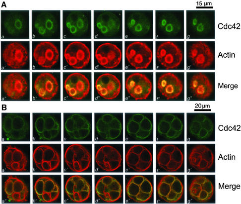
Cdc42 is primarily located to the apical plasma membrane of resting and secreting gastric parietal cells. Cultured gastric parietal cells were prepared as described in MATERIALS AND METHODS. The cultured cells were either maintained in 100 μM cimetidine for resting (A) or treated with 100 μM histamine plus SCH28080 (B) for 20 min before fixation. Fixed and permeabilized cells were incubated with a Cdc42 mAb followed by an FITC-conjugated goat anti-mouse IgG. The cells were also counterstained with rhodamine-labeled phalloidin to visualize filamentous actin cytoskeleton of the cells. Individual fluorescent images from two fluorophores were collected and presented using Adobe Photoshop. (A) This triple montage represents a series of optical sections, spaced ∼0.4 μm in the z-axis, from the bottom (a, a′ and a") to the top (g, g′, and g"), of a resting parietal cell doubly stained for Cdc42 (green, a–g), F-actin (red, a′–g′), and their merges (a"–g"). Cdc42 is chiefly localized to the intracellular of parietal cells, with a distribution profile similar to that F-actin, except for the basolateral surface staining of F-actin. Bar, 15 μm. (B) This triple montage of a series of optical sections was arranged in a similar way to that of A, but in this case the parietal cells were stimulated with histamine. Cdc42 remains localized to the apical plasma membrane of secreting cells in addition to cytoplasmic staining. The distribution of Cdc42 to apical plasma membrane is superimposed to that of F-actin staining. Bar, 20 μm.

