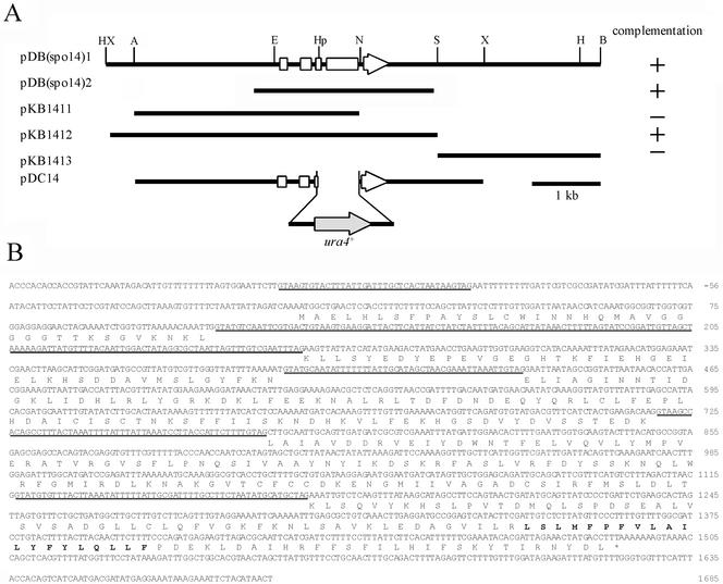
Figure 1.
Cloning, disruption, and the nucleotide sequence of the spo14+ gene. (A) Restriction map, subcloning, and disruption of the spo14+ gene. Plasmids pDB(spo14)1 and pDB(spo14)2 were independently isolated. The white arrow indicates the region and direction of the spo14+ ORF. Restriction enzyme sites: A, ApaI; B, BamHI; E, EcoRI; H, HindIII; Hp, HpaI; N, NsiI; S, SacI; X, XbaI. (B) Nucleotide sequence of spo14+ and its predicted amino acid sequence. Putative introns are underlined. A region indicated by bold letters is a hydrophobic stretch of 20 amino acid residues.

