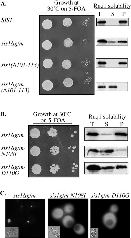
Figure 5.
Unique residues within the G/F region of Sis1 are essential for the maintenance of [RNQ+] in the absence of the G/M region. (A and B) Left, 10-fold serial dilution series of cell suspensions from Δsis1 cells carrying SIS1 on a URA3-based plasmid, as well as a second plasmid expressing wild-type or the indicated mutant were grown on medium containing 5-FOA for 3 d at 30°C. Right, Rnq1 aggregation assays were performed using lysates from Δsis1 cells expressing the specified sis1 mutants. Equivalent amounts of total lysate (T), supernatant (S), and pellet (P) fractions from aggregation assays of lysates from the indicated strains were subjected to SDS-PAGE and analyzed for the presence of Rnq1 by immunoblot analysis. (C) Fluorescence microscopy of Rnq1-GFP in Δsis1 expressing sis1Δg/m, sis1Δg/m-N108I, or sis1Δg/m-D110G as described in Figure 2.

