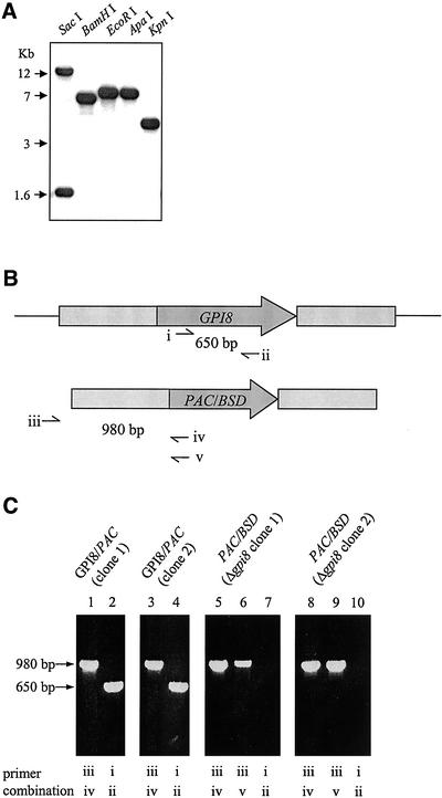
Figure 2.
Generation of Δgpi8 mutants (A) Southern blot analysis of the T. brucei GPI8 gene. DNA was cut with SacI, BamHI, EcoRI, ApaI, or KpnI, transferred onto nylon membrane and hybridized with the ORF of the gene. (B) Schematic representation of the GPI8 locus (top) and PAC/BSD replacement construct (bottom) with open reading frames (dark gray) and GPI8 flanking regions (light gray) used in the integration construct. Thin lines represent regions not present in the integration construct. Primer sites used to confirm presence of GPI8 (i and ii) and correct integration of PAC/BSD constructs (iii with iv or v) are indicated. (C) PCR of genomic DNA to determine correct integration of markers and deletion of GPI8. GPI8/PAC (clone 1) and (clone 2) are two independent heterozygotes containing a PAC (lanes 1 and 3) and a GPI8 (lanes 2 and 4) gene. Derived from these are two independent Δgpi8 clones, containing PAC (lanes 5 and 8) and BSD (lanes 6 and 9), but lacking GPI8 (lanes 7 and 10). Δgpi8 clone 1 was used for all subsequent studies. Some results were confirmed with clone 2 (unpublished data).

