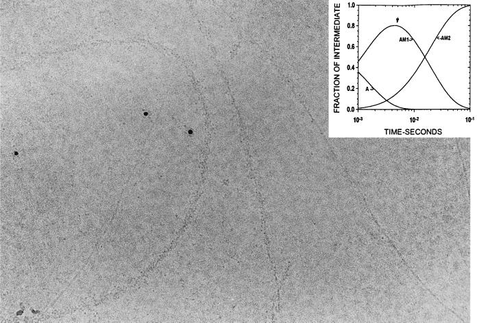
Figure 3.
Electron micrograph of actin sprayed with S1 5 ms before freezing. The S1 bound to the actin is disordered. There is also some S1 visible in the background that has not bound to actin filaments, as well as bare actin filaments. The characteristic subunit and helical substructure is clearly visible in the bare actin filaments. The six intensely black round particles are 5-nm colloidal gold that was included in the spray to aid in searching for areas of the grid that had received spray droplets. Some clustering of the S1 around the gold particles was seen; this depended on the particular batch of gold used and probably resulted from incomplete coverage of the BSA coupled to the gold. The insert shows the time course of the reaction intermediates calculated from the mechanism in Eq. 2 with an arrow indicating the time of freezing. Experimental conditions: 5 mM Mops/2.0 mM MgCl2/0.5 mM DTT, pH 7.0, 4°C. (×388,000.)

