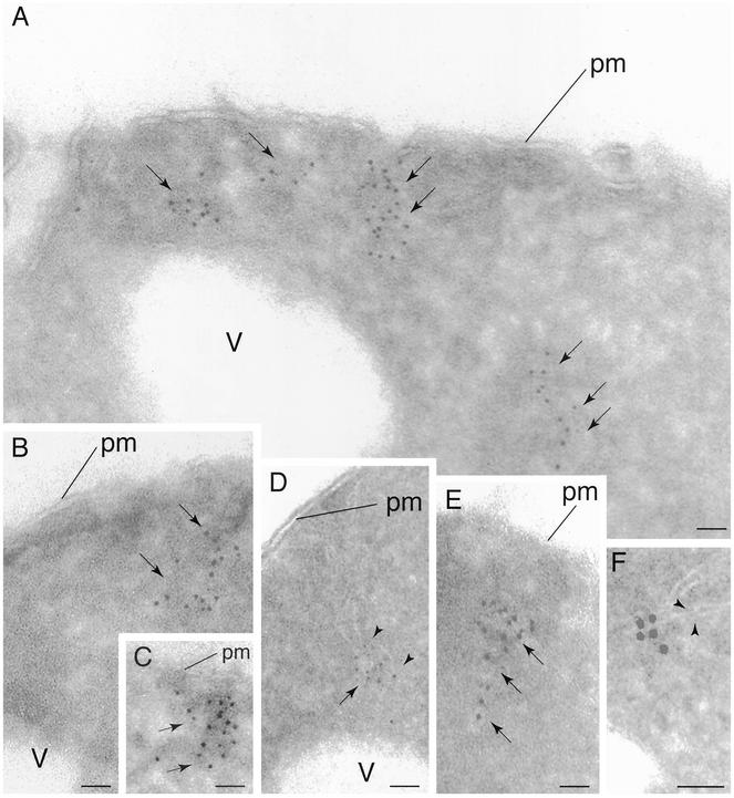
Figure 3.
Localization of Tor2p by IEM. Cells expressing HA3-Tor2p were grown in rich media (YPD) and prepared for electron microscopy and probed with anti-HA antibody, followed by incubation with secondary antibody decorated with 5-nm gold particles. In A–F, arrows denote regions where gold particles are clustered. In D and F, arrowheads point out regions where membrane tracks are clearly evident. pm, plasma membrane; v, vacuole. Bar, 85 nm.

