Figure 3.
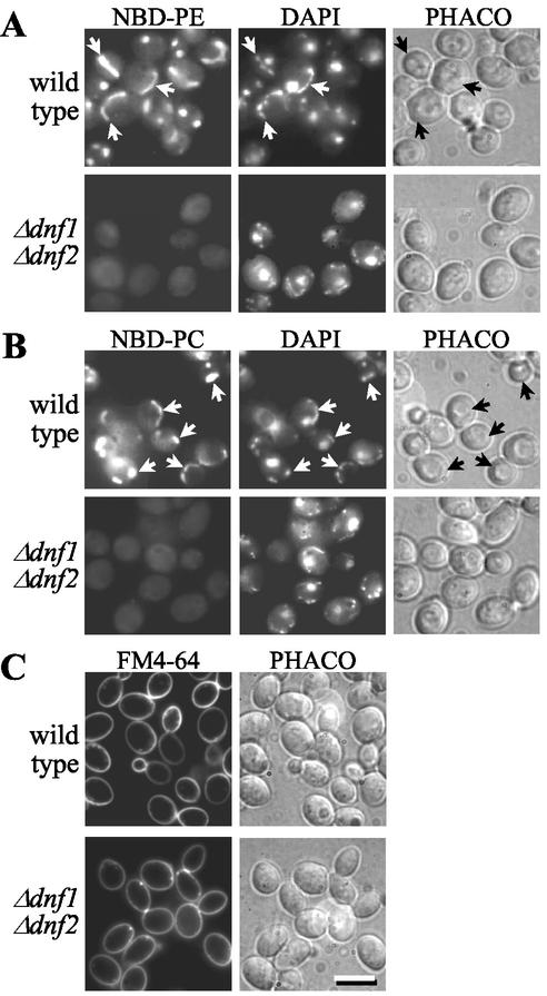
Δdnf1Δdnf2 cells are defective in the nonendocytic uptake of NBD-PE and -PC. Wild-type and Δdnf1Δdnf2 cells, prestained with 1 μg/ml DAPI for 30 min at 30°C, were incubated for 60 min at 2°C with 100 μM NBD-lipid (A and B) or 40 μM FM4-64 (C) before imaging by fluorescence (NBD, DAPI, FM4-64) or phase contrast (PHACO) microscopy. Note that wild-type cells accumulate NBD lipids primarily in mitochondria, as indicated by colocalization with DAPI fluorescence (arrows). For each NBD lipid examined, TLC analysis revealed that >80% of cell-associated NBD fluorescence corresponded to intact lipid. Bar, 10 μm.

