Figure 6.
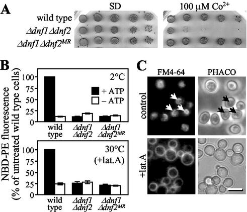
Metal sensitivity of Δdnf1Δdnf2 cells is secondary to the defect in NBD-lipid uptake. (A) Serial fivefold dilutions of wild-type, Δdnf1Δdnf2 and metal-resistant Δdnf1Δdnf2 cells (Δdnf1Δdnf2 MR) were spotted onto SD plates or SD plates containing 100 μM CoCl2. Plates were scanned after 3 d of incubation at 30°C. (B) Cells, preincubated in SD medium (+ATP) or SSA medium (−ATP), were labeled with 100 μM NBD-PE at 2 or 30°C and then analyzed by flow cytometry as in Figure 5. For labeling at 30°C, preincubation was with 20 μM latrunculin A (+lat. A). NBD-PE uptake was expressed as percentage fluorescence intensity relative to control wild-type cells (+ATP). Results represent the means ± SEM of three independent experiments. (C) Wild-type cells, preincubated in SD medium (control) or SD medium containing 20 μM latrunculin A (+lat. A) for 45 min at 30°C, were labeled with 40 μM FM4-64 for 30 min at 30°C and then imaged as in Figure 3. Arrows mark labeled vacuolar membranes. Bar, 10 μm.

